Kidney Stone & Endourology Care in Aligarh

Kidney Stones: Symptoms, Diagnosis & Treatment Options

Kidney stones can cause sudden severe pain, blood in urine, nausea, urinary symptoms or infection, although some stones are discovered incidentally. Dr. Parwez Alam provides evidence-based evaluation and treatment planning for kidney and ureteric stones, including minimally invasive endourology procedures when clinically appropriate.

Kidney & Ureteric Stone Evaluation

Clinical assessment and imaging-led planning.

RIRS • URSL • PCNL

Modern endourology options when clinically appropriate.

Personalised Treatment Planning

Choice based on stone and patient factors.

Prevention & Follow-Up Guidance

Recurrence risk and follow-up considered after treatment.
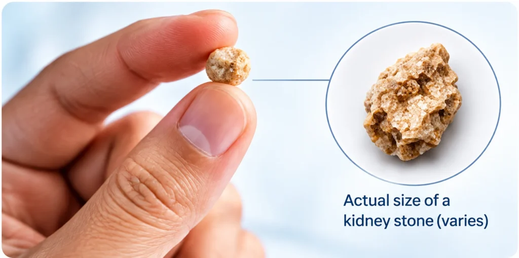
What Are Kidney Stones?

Kidney stones are solid deposits that form from substances in urine. A stone may remain inside the kidney or move into the ureter, the tube that drains urine from the kidney to the bladder. A stone that moves into the ureter may block urine flow and cause renal colic, which is typically sudden, severe pain in the side or back that may radiate towards the groin. Not every stone causes symptoms, and not every stone requires a procedure. The appropriate plan depends on the stone’s size, position and composition, whether it is causing obstruction or infection, the condition of the kidney, previous stone history and the patient’s overall clinical situation.

Kidney Stone

A stone located within the kidney. It may be symptomatic or found incidentally on imaging.

Ureteric Stone

A stone that has moved into the ureter and may cause colicky pain, obstruction or urinary symptoms.

Single or Multiple Stones

Some patients have one stone, while others may have stones in both kidneys or recurrent episodes over time.

Recurrent Stone Disease

Repeated stone formation may justify additional metabolic evaluation and prevention planning based on individual risk.

Common Symptoms of Kidney or Ureteric Stones

Severe Flank / Side Pain

Sudden pain in the side or back, often coming in waves.

Pain Radiating to Groin

Pain may travel towards the lower abdomen or groin.

Blood in Urine

Urine may appear blood-stained or blood may be detected on testing.

Nausea / Vomiting

Severe stone pain can be associated with nausea or vomiting.

Urinary Symptoms

Burning, frequency or urgency can occur, especially with lower ureteric stones.

Reduced Urine / Difficulty Passing Urine

Difficulty passing urine or reduced urine output can occur in selected situations.

Causes and Risk Factors Can Differ Between Patients

Stone formation is influenced by the concentration of substances in urine and by individual metabolic, dietary, anatomical, medical and genetic factors. In many patients, more than one factor contributes.

Low Fluid Intake / Concentrated Urine

Insufficient fluid intake or high fluid loss can lead to more concentrated urine and may increase stone risk.

Previous or Family History

People who have formed stones before, or who have a family history of stone disease, may have a higher risk of recurrence.

Dietary & Metabolic Factors

Dietary patterns, excess salt intake and selected metabolic abnormalities can influence stone formation in susceptible individuals.

Urinary Infection

Certain infection-related stones can develop in association with urinary tract infection.

Medical / Anatomical Factors

Selected medical conditions, urinary tract abnormalities or problems with urine drainage can contribute to stone formation.

Medicines & Genetic Conditions

Some medicines and uncommon inherited disorders can contribute to specific stone types and may require targeted evaluation.

How Kidney Stones Are Evaluated

Evaluation usually begins with symptoms, medical history, examination and review of previous reports. Imaging is used to confirm the presence, size and location of a stone and to assess whether urine drainage is obstructed. Urine and blood tests may be needed to look for infection, kidney function changes and selected metabolic factors.

Clinical Assessment

Symptoms, previous stone episodes, medicines, medical conditions, previous procedures and relevant family history are reviewed.

Urine Testing

Urinalysis and, when indicated, urine culture can help assess blood in urine and possible infection.

Blood Tests

Kidney function and selected blood parameters may be checked depending on the clinical situation.

Imaging

Ultrasound, X-ray or non-contrast CT may be used according to the patient, urgency and clinical question. CT is particularly useful for defining stone size, location and density in many adult patients.

Stone Analysis / Metabolic Evaluation

If a stone is recovered, analysis may help identify its composition. Selected recurrent or high-risk patients may need additional metabolic evaluation, including urine studies.

Treatment Depends on the Stone and the Patient

No. Some small stones, particularly selected ureteric stones, may pass without surgery when pain is controlled and there is no infection, threatening obstruction or other reason for urgent intervention. Observation requires appropriate follow-up because symptoms alone do not always confirm whether a stone has passed.

Active Treatment May Be Considered When

✓ The stone is unlikely to pass spontaneously
✓ Pain remains significant or recurrent despite appropriate treatment
✓ There is persistent obstruction or concern about kidney function
✓ Infection is associated with obstruction
✓ The stone is growing or causing repeated clinical problems
✓ The patient’s anatomy, occupation, travel needs or other clinical circumstances favour active treatment
✓ A large or complex renal stone is present

From Observation to Minimally Invasive Stone Removal

Observation & Symptom Control

Selected stones can be monitored when spontaneous passage is reasonable and there is no urgent indication for intervention. Follow-up is important.

Shock Wave Lithotripsy (SWL)

Shock waves can fragment selected stones from outside the body. Effectiveness depends on stone size, location, density, anatomy and equipment availability.

RIRS

Flexible ureteroscopy is used to reach selected stones inside the kidney through the natural urinary passage without a skin incision.

Medical Expulsive Therapy

Ultrasound, X-ray or non-contrast CT may be used according to the patient, urgency and clinical question. CT is particularly useful for defining stone size, location and density in many adult patients.

URSL / Ureteroscopy

An endoscope is passed through the urinary tract to reach and treat selected ureteric stones, often using laser fragmentation where appropriate.

PCNL

A keyhole tract is created through the back to access the kidney. PCNL is commonly used for larger or complex renal stones and is a guideline-supported first-line option for many stones larger than 2 cm. [Link: /pcnl/]

Urgent Drainage

When an obstructed kidney is infected or urgent decompression is otherwise required, a ureteric stent or nephrostomy may be used before definitive stone treatment.

Individualised Planning

Stone location, size, density, anatomy, infection, kidney function, blood-thinning medicines, previous treatment and patient preference all influence the treatment plan.

RIRS vs URSL vs PCNL

URSL

Often used for selected ureteric stones. The ureteroscope reaches the stone through the urinary passage; fragmentation and/or extraction may be performed.

RIRS

Flexible endoscopic access to the kidney through the urinary tract. Commonly considered for selected renal stones where this approach is clinically suitable.

PCNL

Percutaneous keyhole access directly into the kidney. Commonly used for larger, complex or high-burden renal stones; EAU guidance supports PCNL as first-line treatment for renal stones >2 cm.

Which is right?

There is no single “best” procedure for every stone. Selection depends on stone and patient factors, expected clearance, procedural risks, need for staged treatment and local expertise.

Before & After a Stone Procedure

Before Treatment

Review imaging and symptoms; check urine for infection when indicated; review blood tests and kidney function when required; discuss medicines including blood thinners; explain anaesthesia, procedure-specific risks and expected recovery; discuss whether a temporary ureteric stent may be required.

After Treatment

Follow prescribed medicines and hydration advice; recovery instructions differ by procedure; temporary urinary symptoms can occur after instrumentation or stent placement; seek medical advice for fever, worsening pain, inability to pass urine or other concerning symptoms; attend follow-up and discuss stone analysis/prevention when relevant.

Prevention Starts with Understanding Your Stone Risk

After an acute stone episode is managed, prevention becomes important because some patients develop recurrent stones. General measures often include maintaining adequate fluid intake and reviewing dietary and metabolic risk factors. Prevention should be adapted to the stone type, recurrence pattern, medical history and metabolic findings where available.

Hydration

Maintain regular fluid intake through the day unless a clinician has advised fluid restriction for another medical condition.

Balanced Diet

Avoid extreme “stone diets.” Prevention should be based on the stone type and individual metabolic findings where available.

Salt Awareness

High salt intake can influence urinary calcium handling; moderation may be advised as part of an individual prevention plan.

Stone Analysis

If a stone is available, laboratory analysis can help guide stone-specific prevention.

Metabolic Evaluation

Recurrent or high-risk stone formers may benefit from additional blood and urine assessment.

Follow-Up

Imaging and clinical follow-up may be recommended depending on residual stones, recurrence risk and the treatment performed.

Kidney Stones in Special Situations

Pregnancy

Evaluation and treatment require special attention to imaging and medication safety. Ultrasound is generally preferred as first-line imaging.

Children

Stone disease in children may require paediatric-specific imaging, metabolic evaluation and treatment planning.

Single Functioning Kidney

Symptoms or obstruction may need more urgent assessment because preserving drainage and kidney function is especially important.

Recurrent Stones

Repeated stone formation may justify stone analysis, metabolic assessment and a structured prevention plan.

Urinary Infection

Fever or infection with obstruction requires urgent assessment; definitive stone treatment may need to follow drainage and infection control.

Blood-Thinning Medicines

Procedure selection and timing may be affected by anticoagulant or antiplatelet therapy. Medicines should not be stopped without medical advice.

A Structured Approach to Kidney Stone Evaluation and Treatment

Dr. Parwez Alam’s kidney stone practice focuses on accurate evaluation, clear discussion of treatment choices and selecting an endourology approach according to the individual clinical situation. Where a procedure is required, options may include URSL, RIRS or PCNL depending on stone location, size, complexity, anatomy and other patient factors.

Review Symptoms & Reports

Previous imaging, laboratory tests, prescriptions, procedure records and current symptoms are reviewed.

Confirm the Clinical Problem

Stone location, burden, obstruction, infection risk and kidney function are assessed using appropriate investigations.

Discuss Options

Observation, medical management, SWL or endourological treatment options are discussed when relevant to the patient’s situation.

Plan Treatment

If intervention is required, the procedure is selected according to expected benefit, risk, anatomy and patient needs.

Recovery & Prevention

Follow-up includes recovery guidance, evaluation of residual/recurrent stones where appropriate and recurrence-prevention advice.

Have Kidney Stone Reports? Start with a Structured Review

Patients outside Aligarh may use online consultation for selected needs such as review of CT/ultrasound reports, discussion of an existing diagnosis, second-opinion questions, post-treatment follow-up or guidance before planning an in-person visit. Imaging itself may still need to be reviewed directly, and some symptoms or clinical situations require urgent or in-person assessment.

Report Review

Share available imaging reports, laboratory results, discharge summaries and previous procedure details.

Second Opinion

Discuss a proposed stone procedure and understand why a particular approach may have been recommended.

Pre-Visit Planning

Clarify which reports and records may be useful before travelling to Aligarh for assessment.

Follow-Up

Selected recovery and report-review discussions may be suitable for online follow-up when clinically appropriate.

Frequently Asked Questions

Can a kidney stone pass on its own?
Some smaller stones may pass spontaneously, particularly when located in the ureter, but the likelihood depends on size, location and individual anatomy. Observation is appropriate only when there is no urgent reason for intervention and follow-up is arranged.
Seek urgent assessment for fever or chills, severe uncontrolled pain, persistent vomiting, markedly reduced urine output, concern about infection, or symptoms in a person with a single functioning kidney or other high-risk situation.
The appropriate imaging depends on age, pregnancy status, urgency and the clinical question. Non-contrast CT is highly informative in many adults with suspected renal colic, while ultrasound is preferred first-line in pregnancy and children.
No. Some stones can be observed or managed medically. Intervention is considered when spontaneous passage is unlikely or when there is persistent pain, obstruction, infection, stone growth, repeated problems or other clinical indications.
URSL is commonly used to treat selected ureteric stones; RIRS uses flexible endoscopic access to treat selected stones inside the kidney; PCNL uses a keyhole tract into the kidney and is commonly used for larger or complex renal stones.
Is PCNL always required for a large kidney stone?
PCNL is a guideline-supported first-line option for many renal stones larger than 2 cm, but the final decision depends on anatomy, stone complexity, patient health, previous treatment and other clinical factors.
Yes. Recurrence can occur, particularly in people with persistent metabolic, dietary, anatomical or genetic risk factors. Prevention planning may include hydration, stone analysis and metabolic evaluation where appropriate.
Adequate fluid intake is an important prevention measure. EAU guidance recommends fluid intake sufficient to produce more than about 2.5 litres of urine per day for appropriate adults, but people with heart, kidney or other conditions may need individualised advice.
Online consultation may be useful for report review, second opinions and selected follow-up. Severe pain, fever, vomiting, reduced urine output or other urgent symptoms require prompt in-person assessment.
Bring available ultrasound or CT reports/images, urine and blood test results, previous prescriptions, discharge summaries, records of earlier stone procedures, current medicines and any prior stone-analysis results.
hacklink hack forum hacklink film izle hacklinkbetsat girişbetsat girişbetbomfireball casinoporno sitelericasibomcasibom girişjojobetjojobet giriş
Scroll to Top